Some impressive tools are coming online on the University of Saskatchewan campus. While they are intended primarily for human health applications, there is great potential for those same tools to aid in plant research. A new PET-CT scanner is currently being installed at Royal University Hospital. It is scheduled to be available for clinical patients at the end of April. Dr. Paul Babyn, head of the Department of Medical Imaging, says, “Our mandate goes beyond clinical service. The PET-CT is also available for researchers looking for the kind of resolution the scanner can provide.” 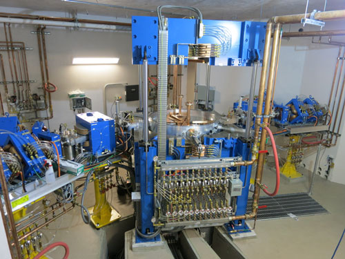
TR-24 cyclotronFor example, the scanner could be used to discover how the body metabolizes plant compounds. Compounds would be tagged and imaged after being consumed by human subjects. We know anthocyanins are anti-oxidants; but do we know how or where the body absorbs them? This kind of research is just waiting to be undertaken.
Most isotopes used in medical imaging (18F, 11C, 13N, 15O) have short half-lives (two minutes to two hours). The PET-CT scanner detects the decay of the isotopes, so imaging needs to take place during that time frame. For very short-lived isotopes, such as 13N and 12C, the imaging would probably have to take place in the same building as the cyclotron.Just up the road from RUH, the Sylvia Fedoruk Canadian Centre for Nuclear Innovation and University of Saskatchewan are working on bringing another imaging tool online: a cyclotron. The cyclotron is a particle accelerator that transforms charged hydrogen ions into radioactive, proton-rich isotopes.
While there are no current plans to include a detector in the building, Ian Swainson, Special Advisor at the Fedoruk Centre, says, “We have space available in the building. We’re hoping a partner will step forward who wants to develop an imaging tool in that space.” There is time for those plans to develop, as the cyclotron’s current construction schedule is about two years. Another piece of the tool, which should be considered as the cyclotron is developed, is having the right detector: a PET-CT scanner is usually built to accommodate a human.
For plant- or animal-related applications, customized detectors would be preferable. For example, in positron autoradiography, detectors are placed directly onto thin tissue, such as a leaf. Positrons can escape the leaf and annihilate within the detector, allowing spatial resolution to a few tenths of a millimeter. PET-CT scans have already demonstrated potential in plant research, with their ability to show real-time soil-plant interactions, as well as real-time photosynthesis activity on the surface of leaves. One of the key benefits of PET-CT is the preservation of the research subject. Many imaging tools destroy the subject in the process of imaging, but with PET-CT, it is possible to return to the same subject later in its life to continue testing.
Martin Reaney, SMA Chair, Lipids Quality & Utilization, and President & CEO of Prairie Tide Chemicals, Inc., can list several research opportunities afforded by the cyclotron and PET-CT scanner. He’s also excited about the possibilities raised by having a cyclotron and synchrotron so close together: “You can get a combination of knowing what is happening, and where, when you combine the imaging and the chemistry. Having the two tools together is an amazing synchronicity.” The cyclotron can increase our knowledge of plants from root to tip.
By tagging certain molecules and imaging their interaction with plants, there is potential to explore photosynthesis up close. By having a better idea of what parts of a plant do the majority of photosynthesis, breeders can draw connections between plant architecture and plant yield. “We can help plants perform as effectively as possible,” says Reaney. While researchers know that plants communicate between roots and leaves constantly, it isn’t yet known how quickly those messages are sent. “How quickly do plants respond to sun going behind a cloud?” Reaney gives as an example. “We would be increasing the resolution of knowledge we have about plants.”
Exploring root systems and their nutrient uptake is another option, as well as environmental impacts on root systems, such as rhizobia. “There is much to learn about how plants interact with commensal or competitive organisms.” While the size of plants and some of their parts can be challenging to image—imagine the specific parts of a flower head, or pollen—there is potential for detecting the vigor of seedlings as well as nutrition traveling to flowers, and measuring results.
All this potential work will contribute to the existing body of knowledge related to plant physiology, and aid plant breeders and agri-business get the optimal performance from plants. Reaney concludes, “We do have knowledge in these areas, but sharper tools will bring more answers.”
Noelle Chorney is a freelance writer in Saskatoon, SK.